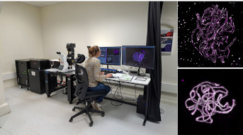
Arrival of a STED high-resolution microscope on the Cytology/Imaging Platform of the IJPB Plant Observatory
Fluorescence microscopy traditionally cannot resolve two points closer than 250 to 300 nm apart. The STED microscope, however, surpasses this resolution limit, providing new possibilities for research teams working at the forefront of current science, particularly in high-resolution subcellular imaging, such as cell wall nanostructures, intracellular vesicles, actin filaments, microtubules, large protein complexes, chromosomes, protein interactions, microenvironmental influences, and detection of multiple probes. It also enables 3D reconstructions and modeling.
The "Cytology/Imaging" plateform PO-Cyto of the Plant Observatory OV has acquired a new microscope, the Leica Stellaris 8, that enables high-resolution imaging using STED (Stimulated Emission Depletion Microscopy) and FLIM (Fluorescence Lifetime Imaging Microscopy) techniques. This acquisition was made possible thanks to multiple funding sources (Sesame, SPS, CNOC, IBISA, INRAE). The Leica Stellaris 8 enables both classic confocal and STED imaging, suitable for both live and fixed samples, such as tissues, cells, and subcellular compartments. The STED mode provides 3D resolution beyond the diffraction limit, enabling better visualization of nanostructures that were previously undetectable with conventional microscopy. Additionally, the FLIM module provides high-speed fluorescence lifetime analysis. This advanced equipment opens new research avenues, particularly in functional imaging, by facilitating the study of molecular interactions and the influence of the microenvironment.
STED is a scanning fluorescence microscopy technique that uses stimulated emission depletion (donut) to remove fluorescence from the outer regions of the excitation optical impulse response. The width of the central region of the donut that can still emit light is adjusted by fluorescence saturation. This nanoscopy technique achieves resolutions close to 50 nm in XY and 100 nm in Z when using the 3D STED option, across a spectral range from 440 to 790 nm. The system features an exclusive design for high-sensitivity spectral excitation and detection, based on a prism with freely adjustable detection bands for all fluorescence channels. In addition, a pulsed white laser (440 to 700nm) combined with sensitive detectors operating in photon-counting mode, enables the FLIM technique. FLIM quantifies the fluorescence lifetime of a fluorophore for each pixel of the image, enabling us to work at very high speed, and thus to study rapid interactions between proteins (FRET-FLIM), protein-ligands or proteins in their microenvironment (biosensors).
As the third facility of its kind in France, this advanced equipment is accesible to the entire scientific community. It will train upcoming researchers on emerging themes, including during a summer school planned for late June 2025 at the IJPB, and will foster new research on nanostructures and their functions. These approaches are essential for a better understanding of complex biological mechanisms and enhancing or refining scientific models.
In connection with the research developed at the Institute Jean-Pierre Bourgin for Plant Sciences.
Back

Legend: Left - Microscope Stellaris 8 STED FLIM at the "Cytology/Imaging" PO-Cyto plateform. Bottom right - Visualization of the chromosomes of an Arabidopsis thaliana meiocyte using confocal. Top right -in super-resolution microscopy. In both cases, after immunostaining for two proteins: REC8 (gray signal), revealing the chromosome axes, and ZYP1 (magenta signal), marking the synaptonemal complex (SC). At this stage of meiosis, homologous chromosomes are closely paired along their entire length. Super-resolution microscopy shows that the chromosomes are aligned at a constant distance of 200 nm, with the SC positioned between them.
IJPB and BAP (Plant Biology and Breeding) INRAE Division Highlight
Contacts:
Bertrand Dubreucq, contact
Alice Vayssières, contact
Samantha Vernhettes, contact
IJPB team:
The "Cytology/Imaging" plateform PO-Cyto